ECG Reading and Interpretation for NCLEX-RN/NMC CBT Exam Prep.
You look at the EKG rhythm strip. Something is clearly wrong, and wonder, “What should the nurse do first?” while the four options all seem possible.
That is exactly where most candidates lose marks on ECG questions. When they can name the rhythm, sometimes they skip what the examiner is really testing- What this rhythm means for this patient, right now.
Key Takeaways: ECG Interpretation
• Assess the patient first. A strip never tells you the whole story.
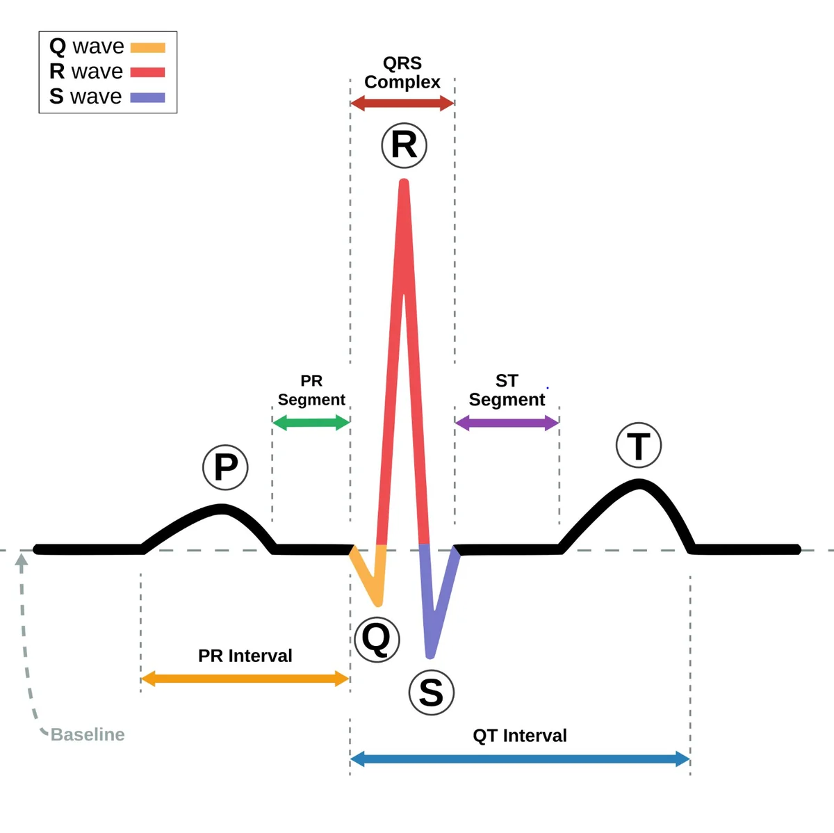
• Read systematically: rate, rhythm, P waves, PR interval, QRS width.
• Pulse or no pulse changes everything, especially for ventricular tachycardia.
• Shockable arrest rhythms: ventricular fibrillation (VF) and pulseless ventricular tachycardia (pVT).
• Non-shockable arrest rhythms: pulseless electrical activity (PEA) and asystole.
• Unstable with a pulse usually means synchronised cardioversion (tachycardia) or atropine, then pacing (bradycardia).
Important Insight: Recognise the rhythm. Assess the patient. Choose the priority action. Evaluate the response.
1. How to Read an ECG Strip: The 5-Step Method
Quick answer: Work through rate, rhythm, P waves, PR interval and QRS width in that order. Do not name the rhythm from one feature. Naming it too early is the most common reason candidates pick a wrong answer.
An electrocardiogram (ECG, also called EKG) records the heart’s electrical activity. For patient, NHS explains how ECG testing works provide a clinical guidance. For exam prep, use this routine:
Step 1: Rate
Normal adult sinus rate is 60 to 100 beats per minute. For an irregular rhythm, count the QRS complexes in a six-second strip and multiply by 10. For a regular rhythm at standard speed, 300 divided by the number of large squares between R waves gives a quick estimate.
Example: Seven QRS complexes in six seconds = about 70 beats per minute.
Step 2: Rhythm
Compare the R–R intervals. Consistent spacing is regular. Varying spacing is irregular. An irregularly irregular pattern, with no repeating pattern at all, should make you think of atrial fibrillation.
Step 3: P waves
Ask three questions: Are P waves present? Do they look the same? Is every P wave followed by a QRS?
Rem. P waves show atrial depolarisation, and their relationship with the QRS reveals conduction problems.
Step 4: PR interval
Normal is 0.12 to 0.20 seconds, which are three to five small squares. Longer than 0.20 seconds suggests first-degree AV block.
A PR that keeps lengthening, or a QRS that suddenly drops, points to a higher-grade block.
Step 5: QRS width
Under 0.12 seconds is narrow, while 0.12 seconds or more is wide.
A wide-complex tachycardia of uncertain origin is treated as ventricular until confirm
cue: RATE → RHYTHM → P WAVES → PR → QRS.
Worked example
A six-second strip shows seven QRS complexes with uneven spacing, no clear P waves and narrow QRS complexes. Rate about 70 and Rhythm irregularly irregular. There are no P waves and PR not measurable. As weel, QRS is narrow.
Interpretation: atrial fibrillation with a controlled ventricular rate.
Exam tip-under exam pressure, your eye will jump to the most dramatic feature on the strip. Use the same five steps every time, even on questions that look easy. The routine is what protects you from the distractors.
2. Ensure Patient Stability
An abnormal rhythm does not automatically need a shock or an emergency drug. The deciding question is whether the rhythm is causing adverse features: shock, syncope, myocardial ischaemia or heart failure.
Look for hypotension, cold clammy skin, new confusion, fainting, ischaemic chest pain, and breathlessness with crackles. Then establish whether the rhythm is the cause.
Quick scenario
Patient A: heart rate 52/min, feels well, blood pressure normal. Patient B: heart rate 42/min, confused, blood pressure 76/40 mmHg. Which patient should be prioritised?
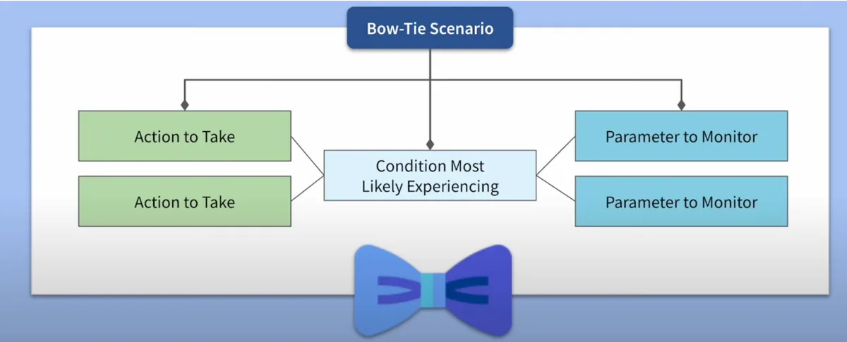
Patient B. The patient number is not the point, their poor perfusion is. This is the reasoning NCLEX clinical judgement questions reward, and our NCLEX bowtie question guide shows how to connect cues, a condition, actions and monitoring in one chain.
The stable vs unstable cases
|
Situation |
Stable (no adverse features) |
Unstable (adverse features) |
No pulse |
|
Bradycardia |
Observe, find the cause, monitor |
Atropine, then pacing or infusion, call for help |
Cardiac arrest: CPR |
|
Narrow-complex tachycardia |
Vagal manoeuvre, then adenosine |
Synchronised cardioversion |
Cardiac arrest: CPR |
|
Wide-complex tachycardia (VT) |
Expert help, antiarrhythmic drug |
Synchronised cardioversion |
CPR and defibrillation |
|
Atrial fibrillation |
Rate or rhythm control, stroke risk review |
Synchronised cardioversion |
Cardiac arrest: CPR |
For clinical decisions advanced decisions, use the applicable guideline, such as the American Heart Association adult advanced life support guidance or the Resuscitation Council UK 2025 guidelines.
3. Rhythm Mastery
Quick answer: Use this table for fast recall. Each row gives the key ECG clue, the first priority and the trap examiners build into the distractors. Learn the reasoning in the sections below and use this page for last-minute revision.
|
Rhythm |
Key ECG clue |
First priority |
Exam trap |
|
Sinus bradycardia |
Rate below 60, regular, P before every QRS |
Assess perfusion; treat only if unstable |
Atropine for every rate below 60 |
|
Atrial fibrillation |
Irregularly irregular, no P waves |
Check stability, rate or rhythm control, stroke risk |
Anticoagulating without a risk assessment |
|
Atrial flutter |
Sawtooth waves; often about 150 with 2:1 conduction |
As AF; cardiovert if unstable |
Mistaking it for sinus tachycardia |
|
Narrow-complex tachycardia (SVT) |
Regular, 150 to 250, narrow QRS, sudden onset |
Stable: vagal manoeuvre then adenosine. Unstable: synchronised cardioversion |
Adenosine before checking stability |
|
First-degree AV block |
PR above 0.20 s; every P conducts |
Monitor; review medicines |
Treating it as an emergency |
|
Mobitz I (Wenckebach) |
PR lengthens, then a QRS drops |
Monitor; often benign |
Confusing it with Mobitz II |
|
Mobitz II |
Constant PR, sudden dropped QRS |
Close monitoring, pads on, urgent escalation |
Expecting atropine to fix it |
|
Third-degree (complete) block |
P waves and QRS unrelated; slow ventricular rate |
Urgent escalation and pacing |
Reading the P waves as “dropped beats” |
|
PVCs |
Early, wide QRS, no preceding P |
Check symptoms, potassium, magnesium, oxygenation |
Treating every PVC with a drug |
|
VT with a pulse |
Regular wide-complex tachycardia |
Stable: expert help. Unstable: synchronised cardioversion |
Forgetting to check the pulse |
|
Torsades de pointes |
Twisting polymorphic VT, long QT |
Unstable or pulseless: shock. Stable: magnesium |
Synchronising a shock for polymorphic VT |
|
Ventricular fibrillation |
Chaotic, no identifiable QRS |
CPR and defibrillation |
Giving drugs before the first shock |
|
Pulseless VT |
Wide tachycardia, no pulse |
CPR and defibrillation |
Treating it as VT with a pulse |
|
PEA |
Organised rhythm, no pulse |
CPR, adrenaline, reversible causes |
Defibrillating |
|
Asystole |
Flat line (confirm on two leads) |
CPR, adrenaline, reversible causes |
Shocking a flat line |
4. Slow Rhythms: Sinus Bradycardia and Heart Blocks
Sinus bradycardia: treat the patient, not the number
Quick answer: Sinus bradycardia is a sinus rhythm below 60 beats per minute. It needs treatment only if it is causing poor perfusion. A well patient at 52/min needs monitoring, not atropine.
What it looks like: rate below 60, regular rhythm, a normal P wave before every QRS, constant PR interval and usually a narrow QRS.
Stable patient: continue monitoring, look for causes (beta-blockers, hypoxia, raised intracranial pressure, inferior myocardial infarction, or hypothyroidism) and report according to local protocol.
Unstable patient: support airway and breathing, give oxygen if hypoxic, attach monitoring, secure IV access, record a 12-lead ECG and follow your bradycardia algorithm.
The AHA algorithm describes clinically significant bradycardia as typically below 50/min with symptoms, which is the reason exams stress perfusion over the number.
UK vs US: Atropine and escalation
|
|
US (AHA) |
UK (Resuscitation Council UK) |
|
First dose |
Atropine 1 mg IV |
Atropine 500 micrograms IV |
|
Repeat |
Every 3 to 5 minutes |
Every 3 to 5 minutes |
|
Maximum total |
3 mg |
3 mg |
|
If atropine fails |
Transcutaneous pacing, or epinephrine or dopamine infusion, and expert help |
Transcutaneous pacing, or isoprenaline or adrenaline infusion, and expert help |
|
Cautions |
Avoid relying on atropine in heart-transplant recipients and in high-grade block with a wide QRS |
Atropine may be ineffective or unsafe in the same situations; go straight to pacing |
UK guidance also highlights risk of asystole, and recommend pacing readiness even if atropine works:
Recent asystole, Mobitz II block, complete heart block with a wide QRS, and a ventricular pause longer than three seconds do. Review the current Resuscitation Council UK recommendations for the full algorithm.
Why atropine works: it blocks parasympathetic (vagal) effects on the heart, so the sinus node fires faster and AV conduction improves.
However, itt cannot help when the block sits below the AV node or when the heart is denervated after transplant.
Exam cue: SLOW RATE + POOR PERFUSION = ACT NOW.
SLOW RATE + WELL PATIENT = MONITOR.
Heart blocks: Master with three-question method
Do not memorise names first. For every strip, ask:
1. Is every P wave followed by a QRS?
2. Is the PR interval changing?
3. Do the P waves and QRS complexes relate to each other at all?
|
Block |
PR interval |
P and QRS relationship |
Risk |
Nursing priority |
|
First-degree |
Constant, above 0.20 s |
Every P conducts |
Low |
Monitor, review medicines |
|
Mobitz I |
Lengthens until a QRS drops |
Grouped beating |
Low to moderate |
Monitor, report new or symptomatic change |
|
Mobitz II |
Constant on conducted beats |
Sudden dropped QRS |
High |
Pads on, urgent escalation, prepare for pacing |
|
Third-degree |
No relationship |
P and QRS independent |
High |
Urgent escalation, pacing |
Cues:
First-degree: LONG PR, NOTHING DROPS.
Mobitz I: LONGER, LONGER, DROP.
Mobitz II: CONSTANT PR, SUDDEN DROP, DANGER.
Third degree: P AND QRS WORK INDEPENDENTLY.
Why Mobitz II is the dangerous one: the conduction system fails below the AV node and can progress suddenly to complete heart block. In third-degree block, the ventricles rely on an escape pacemaker, typically about 20 to 40/min if ventricular and 40 to 60/min if junctional, which may be too slow to maintain output. Permanent pacing is generally indicated for acquired complete block once reversible causes are excluded.
Examiner trap: Giving atropine for Mobitz II or complete block with a wide QRS and expecting it to work instead of Pacing.
5. Fast Narrow Rhythms: AF, Atrial Flutter and Narrow-Complex Tachycardia (SVT)
Atrial fibrillation vs atrial flutter
Quick answer: Atrial fibrillation (AF) is irregularly irregular with no distinct P waves. Atrial flutter shows sawtooth flutter waves and is often regular, with a ventricular rate of about 150/min when conduction is 2:1.
|
Feature |
Atrial fibrillation |
Atrial flutter |
|
Rhythm |
Irregularly irregular |
Regular if conduction is fixed; irregular if variable |
|
Atrial activity |
No distinct P waves |
Sawtooth flutter waves |
|
Ventricular rate |
Variable |
About 150/min with 2:1 conduction |
|
Main concern |
Stroke from left atrial appendage thrombus, plus rate control |
Same stroke risk; rate often hard to control |
Why AF matters beyond the heart rate: ineffective atrial contraction lets blood pool in the left atrial appendage, which raises the risk of thrombus and stroke. As a result, the nurse thinks about stroke prevention as well as rate.
Stable patient: assess symptoms, control the ventricular rate or restore sinus rhythm as prescribed, and review stroke and bleeding risk. NICE guideline NG196 covers diagnosis, rate control and anticoagulation in detail.
Unstable patient-shock, ischaemia, heart failure, or syncope with synchronised cardioversion. When AF has lasted more than 48 hours or the onset is unknown, cardioversion carries a stroke risk in a stable patient, so teams plan anticoagulation or imaging first.
cue: AFIB = IRREGULARLY IRREGULAR + STROKE RISK.
Examiner trap: Anticoagulation is not automatic for every AF patient. Stroke risk, bleeding risk and contraindications should be taken into account. Our bowtie guide works through a fully solved AF example.
Narrow-complex tachycardia (SVT): When do you administer adenosine?
Quick answer: In a stable patient with a regular narrow-complex tachycardia, try vagal manoeuvres first, then adenosine if they fail.
In an unstable patient, go straight to synchronised cardioversion. Recent AHA material uses the term narrow-complex tachycardia in place of SVT, so you may see both.
Picture it: sudden palpitations, heart rate 190/min, regular rhythm, narrow QRS, P waves hard to see, abrupt onset and termination.
Step 1: Assess stability.
Step 2, if stable: vagal manoeuvres for example the modified Valsalva.
Step 3: adenosine when appropriate.
Unstable patients need urgent synchronised cardioversion, with sedation where the patient is conscious.
|
|
US (AHA) |
UK (Resuscitation Council UK) |
|
Adenosine sequence |
6 mg rapid IV push, then 12 mg if required |
6 mg, then 12 mg, then 18 mg if required |
|
Technique |
Rapid push into a large vein, followed by a saline flush, with continuous ECG monitoring |
Same |
Why adenosine works: it briefly blocks conduction through the AV node, which can break a re-entry circuit and restore sinus rhythm. It will not terminate rhythms that do not depend on the AV node. In atrial flutter or AF it only slows conduction for a moment, which can unmask the underlying atrial rhythm.
Nursing points examiners love: warn the patient about transient chest tightness, breathlessness and flushing; keep the defibrillator nearby; use a reduced starting dose in a transplanted heart or when given through a central line, as per local protocol.
Avoid or seek senior advice in pre-excited AF, second- or third-degree block, sick sinus syndrome and asthma or bronchospasm.
Exam cue: STABLE + REGULAR + NARROW → VAGAL MANOEUVRE → ADENOSINE.
6. Ventricular Rhythms with a Pulse-PVCs, VT and Torsades
Premature ventricular contractions (PVCs)
Quick answer: A PVC is an early beat from the ventricles. It appears as a premature, wide QRS with no preceding P wave. Occasional PVCs are often benign. New, frequent, symptomatic or complex PVCs need assessment and escalation.
Assess symptoms, pulse, blood pressure, oxygenation, potassium and magnesium results and cardiac history.
Escalate for multifocal PVCs, couplets, runs of VT and R-on-T beats.
Exam cue: EARLY + WIDE QRS + NO PRECEDING P = THINK PVC.
Ventricular tachycardia (VT): why the pulse changes your answer
Quick answer: Ask whether the patient has a pulse. VT with a pulse is managed by stability and morphology.
VT without a pulse is cardiac arrest and needs CPR and defibrillation.
|
Situation |
Priority action |
|
Stable monomorphic VT with a pulse |
Monitor, get expert help, prepare a prescribed antiarrhythmic (for example amiodarone; procainamide or sotalol are AHA options) |
|
Unstable monomorphic VT with a pulse |
Synchronised cardioversion (the 2025 AHA update reinforces this for unstable wide-complex tachycardia) |
|
Sustained polymorphic VT |
Treat like VF with an unsynchronised shock, because synchronisation cannot lock on to a changing QRS |
|
Pulseless VT |
High-quality CPR and defibrillation as soon as possible |
Why this matters: VT reduces cardiac output. If the pulse is absent there is no effective circulation and the cardiac arrest pathway applies.
If a pulse is present, stability decides the intervention.
Exam cue: VT + NO PULSE = CPR + DEFIBRILLATION.
Torsades de pointes
Torsades is polymorphic VT linked to a prolonged QT interval. If the patient is pulseless or unstable, give an immediate unsynchronised shock. If the patient is stable, treatment centres on IV magnesium sulfate as prescribed, correcting potassium and magnesium, and stopping QT-prolonging medicines.
Do not delay a needed shock to give magnesium.
7. Cardiac Arrest- VF, Pulseless VT, PEA and Asystole
Quick answer: VF and pulseless VT are shockable.
PEA and asystole are not.
In all four, start high-quality CPR immediately, and treat reversible causes.
|
Rhythm |
Shockable? |
Main intervention |
|
Ventricular fibrillation |
Yes |
CPR and defibrillation |
|
Pulseless VT |
Yes |
CPR and defibrillation |
|
PEA |
No |
CPR, adrenaline (epinephrine), reversible causes |
|
Asystole |
No |
CPR, adrenaline (epinephrine), reversible causes |
Unstable VT with a pulse (synchronised cardioversion) and sustained polymorphic VT (unsynchronised shock) are important emergency electrical decisions, but they are not part of the standard shockable versus non-shockable arrest classification.
The nurse’s first minute
• Confirm the patient is unresponsive and not breathing normally, and call the resuscitation team.
• Start chest compressions: 100 to 120 per minute, 5 to 6 cm deep, full recoil, minimal interruptions, 30 compressions to 2 breaths.
• Attach the defibrillator and analyse the rhythm. Shock if shockable, then resume CPR immediately for two minutes.
• Secure IV or IO access and give adrenaline (epinephrine) according to the algorithm.
• Look for and treat reversible causes (the Hs and Ts below).
UK vs US: drug sequencing in shockable arrest
|
|
UK (Resuscitation Council UK 2025) |
US (AHA) |
|
Adrenaline (epinephrine) |
1 mg after the third shock for VF or pVT, then every 3 to 5 minutes. In non-shockable rhythms, give it as soon as possible. |
1 mg every 3 to 5 minutes. Timing in the shockable pathway was revised in 2025, so check the current algorithm. |
|
Amiodarone |
300 mg after the third shock, then 150 mg after the fifth shock if VF or pVT persists |
300 mg, then 150 mg, for shock-refractory VF or pVT |
|
Alternative |
Lidocaine 100 mg if amiodarone is unavailable |
Lidocaine 1 to 1.5 mg/kg |
Asystole and PEA: why you do not defibrillate
Imagine a patient who has become unresponsive. The monitor shows a nearly flat line and there is no pulse.
Do you defibrillate? No.
Defibrillation terminates shockable ventricular arrhythmias. It cannot restart a rhythm when there is no electrical activity, or when there is organised electrical activity without effective circulation (PEA).
Before declaring asystole: check the leads are connected, the gain is up and the rhythm is confirmed in more than one lead. A loose electrode can mimic a flat line.
Exam cue: ASYSTOLE OR PEA = CPR + ADRENALINE (EPINEPHRINE) + REVERSIBLE CAUSES.
Torsades and VF in arrest: a sustained episode in a pulseless patient is treated as VF, with CPR and defibrillation first.
Reversible causes: UK four Hs and four Ts vs AHA five Hs and five Ts
|
UK: four Hs |
UK: four Ts |
AHA: five Hs |
AHA: five Ts |
|
Hypoxia |
Thrombosis (coronary or pulmonary) |
Hypovolaemia |
Tension pneumothorax |
|
Hypovolaemia |
Tension pneumothorax |
Hypoxia |
Tamponade (cardiac) |
|
Hyper-/hypokalaemia and other metabolic causes |
Tamponade (cardiac) |
Hydrogen ions (acidosis) |
Toxins |
|
Hypo-/hyperthermia |
Toxins |
Hypo-/hyperkalaemia |
Thrombosis (pulmonary) |
|
|
|
Hypothermia |
Thrombosis (coronary) |
In any deteriorating patient, ask which reversible cause best explains what you see.
For NMC CBT revision
Focus on patient safety, recognising deterioration, UK terminology, professional standards and escalation. The NMC publishes its Test of Competence information, and you should check it for the current scope. Our guide to passing the NMC CBT first time covers format, mistakes and study strategy.
In UK practice, NEWS2 supports recognition of deterioration. Know these triggers:
• Aggregate score of 5 or 6: urgent clinical review.
• Aggregate score of 7 or more: emergency-level clinical assessment.
• A score of 3 in any single parameter: urgent clinical review, even if the total is below 5.
Review the scoring system in the Royal College of Physicians’ NEWS2 report. Pair it with ABCDE assessment and SBAR communication.
For NCLEX-RN revision
Practise the six steps of the clinical judgement model: recognise cues, analyse cues, prioritise hypotheses, generate solutions, take action and evaluate outcomes. Our 2026 NCLEX test plan guide explains how that fits the current exam, and our NCLEX-RN preparation guide covers format and strategy.
Coach’s Insight: The skill you are building is clinical judgement, not memorising drug names or ECG patterns. If you can explain why an action comes first, you can answer questions you have never seen.
10. Eight Mistakes That Cost Marks, and a Seven-Day Plan
Eight mistakes to avoid
• Naming the rhythm from one feature. Run all five steps every time.
• Treating the number instead of the patient. Look for adverse features first.
• Giving atropine for every slow rate. Perfusion decides.
• Giving adenosine before checking stability. Unstable patients need cardioversion.
• Missing the pulse check in VT. Pulse changes the whole pathway.
• Synchronising a shock for polymorphic VT or VF. The device cannot sync to chaos.
• Defibrillating PEA or asystole. CPR, adrenaline (epinephrine) and reversible causes.
• Mixing UK and US doses. Match the answer to the guideline the question names.
A seven-day ECG revision plan
Short, repeated sessions beat one long weekend. This plan uses retrieval practice and spaced review, the same methods in our guide to nursing exam memorisation techniques.
|
Day |
Focus |
Task (about 45 minutes) |
|
1 |
Five-step method |
Read 10 strips aloud using rate, rhythm, P, PR, QRS. Write the cue chain from memory. |
|
2 |
Slow rhythms |
Redraw the heart block table from memory. Do Questions 1, 2 and 4. |
|
3 |
Fast narrow rhythms |
Explain AF versus flutter and the adenosine steps aloud. Do Question 3. |
|
4 |
Ventricular rhythms |
Rebuild the VT decision table from memory. Rehearse the pulse question. |
|
5 |
Cardiac arrest |
Say the first-minute checklist aloud. Do Questions 5 and 6. Compare UK and US drug timing. |
|
6 |
Mixed practice |
Do Questions 7 and 8, then 10 new mixed questions. Log every miss with the reason. |
|
7 |
Review and recall |
Re-test only the missed items. Rewrite the 15-rhythm cheat sheet blank-page. |
Self-audit: are you exam-ready on ECGs?
• Can you name the five steps without notes?
• Can you explain, in one sentence, why a stable patient at 52/min does not get atropine?
• Can you state the first action for VT with a pulse and for VT without one?
• Can you list the UK and US atropine and adenosine differences?
• Can you explain why you never shock PEA or asystole?
• Do you reassess after every intervention instead of assuming it worked?
If you answered “not yet” to several, you have found your study plan. That is the point of the audit.
Frequently Asked Questions
What is the easiest way to learn ECG interpretation?
Use the same five steps every time: rate, rhythm, P waves, PR interval and QRS width. Then link each pattern to the patient’s symptoms and the nursing priority. Daily short practice with explanations beats rereading notes.
Is an ECG the same as an EKG?
Yes. Both terms mean electrocardiogram. ECG is the usual UK term, and EKG is common in the United States. The NMC CBT usually uses ECG, and NCLEX materials often use EKG.
How do you read an ECG strip step by step?
Calculate the rate, check whether the rhythm is regular, look for P waves, measure the PR interval (normal 0.12 to 0.20 seconds) and check QRS width (narrow is below 0.12 seconds). Then assess the patient before choosing an intervention.
Which cardiac arrest rhythms are shockable?
Ventricular fibrillation (VF) and pulseless ventricular tachycardia (pVT) are shockable. Treat them with high-quality CPR and defibrillation.
Which cardiac arrest rhythms are non-shockable?
Asystole and pulseless electrical activity (PEA) are non-shockable. Treat them with CPR, adrenaline (epinephrine) and a search for reversible causes.
Do you shock asystole?
No. Asystole is a non-shockable rhythm because defibrillation cannot restart a heart with no electrical activity. First check the leads and gain, then give CPR and adrenaline (epinephrine) and look for reversible causes.
What is PEA?
Pulseless electrical activity means the monitor shows an organised rhythm but there is no effective pulse. It is non-shockable and is managed with CPR, adrenaline (epinephrine) and treatment of the underlying cause.
What is the difference between atrial fibrillation and atrial flutter?
AF is irregularly irregular with no distinct P waves. Atrial flutter shows sawtooth waves and is often regular, commonly around 150 per minute with 2:1 conduction. Both carry stroke risk.
When should a nurse give atropine?
Give atropine when bradycardia causes adverse features such as hypotension, confusion, chest pain or heart failure, and only as prescribed or per protocol. It is not for stable bradycardia. UK first dose is 500 micrograms, US is 1 mg, and both cap the total at 3 mg.
What is the adenosine dose in the UK and the US?
The US (AHA) sequence is 6 mg then 12 mg. The UK (Resuscitation Council UK) sequence is 6 mg, 12 mg, then 18 mg if needed. Give it as a rapid bolus into a large vein with a flush, and always confirm against current guidance.
How do you tell Mobitz I from Mobitz II?
In Mobitz I the PR interval lengthens until a QRS drops. In Mobitz II the PR stays constant and a QRS drops suddenly. Mobitz II is more dangerous because it can progress to complete heart block.
What is the first thing to check in ventricular tachycardia?
Check for a pulse and signs of instability. VT with a pulse is treated by stability: expert help and a drug if stable, synchronised cardioversion if unstable. VT without a pulse is cardiac arrest and needs CPR and defibrillation.
What is the difference between cardioversion and defibrillation?
Synchronised cardioversion times the shock to the R wave and treats unstable tachycardias with a pulse. Defibrillation is an unsynchronised shock for VF, pulseless VT and sustained polymorphic VT.
Do the NMC CBT and NCLEX-RN use the same resuscitation protocols?
No. They share principles but differ in some doses, drug names and timing. The NMC CBT follows UK practice (Resuscitation Council UK), and NCLEX follows US practice (AHA). Learn the guideline for your exam.
Is ECG interpretation on the NMC CBT or NCLEX?
NCLEX-RN can include rhythm strips and cardiac emergencies within its clinical judgement items. For the NMC CBT, expect to recognise common arrhythmias and escalate appropriately, and check the NMC’s current Test of Competence information for exact scope.
Can I pass by memorising ECG rhythms alone?
No. Memorising patterns is not enough. You need to recognise cues, judge stability, choose the priority action and explain the rationale. That is what both exams assess.
Learn the Reasoning, Not Just the Rhythm
If you take one thing from this guide, make it this: ECG questions are rarely about the strip. They are about the patient behind it.
Start with the same five steps. Check for a pulse and adverse features. Pick the safest priority action. Then ask the question the exam is really testing: What should the nurse do next, and why?
Planning to practise abroad as well? Our nursing registration pathways guide compares the UK, Australia and New Zealand, and our NMC OSCE guide explains the practical stage of the UK route.
|
Want a tutor to build your NCLEX-RN or NMC CBT plan with you? Book a free consultation with UKNurses and get matched with a nurse tutor for 1-on-1 coaching built around your weak areas, your schedule and your test date. Prefer to chat? Message the team on WhatsApp. |